The modern smartphone comes with unlimited possibilities for the creative mind. With no more than a few clicks, taps, and pinches of the fingers, anyone can find out practically anything anywhere in the world. But what if we started harnessing the power of our smartphones in our medical schools, on our athletic fields and—more importantly—in developing countries, where patients who need care the most are least likely to receive it? Remarkably, one team has begun doing just that. Using a sophisticated smartphone application in combination with snap-on hardware, a startup named Peek has developed a system for portable eye examinations.
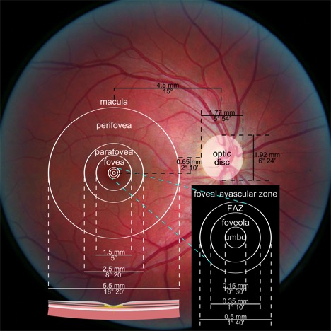
 According to the World Health Organization, 39 million people worldwide are blind, and 246 million have low visual acuity; about 80% of these cases are preventable or curable [1]. When a patient presents with vision impairment, the first thing that is typically done is an examination of the retina, a part of the eye that holds huge amounts of information about the body and its health, with implications for many different branches of medicine. However, analysis of the retina requires a skilled ophthalmologist along with bulky, expensive, and largely immobile diagnostic equipment. With the development of Peek, a person with little training can take high-quality images of the retina anywhere in the world and have those images remotely analyzed by an ophthalmologist [2]. Instead of patients taking long and uncertain trips to the clinic, a single person with a smartphone can travel from village to village, providing comprehensive, high-quality eye examinations for everyone who needs them. Now that we can accurately diagnose patients faster than ever before, we may be able to provide them with the medications, corrective lenses, and treatments they need to live comfortably.
But what if the implications of this technology are even broader? Does Peek have a place in medical schools and clinics? As medical students, we have all looked through the ophthalmoscope and assessed the landmarks of the retina. After the brief exam, we pull out the scope and think, “Wait a minute—was that normal?” With the advent of Peek, students will be able to assess standardized patients, take pictures of their retinas, and consult attending physicians for abnormalities without the need for patients to undergo second exams by attendings. Peek provides a less intrusive, more efficient, and more formative experience that students can learn from exam after exam.
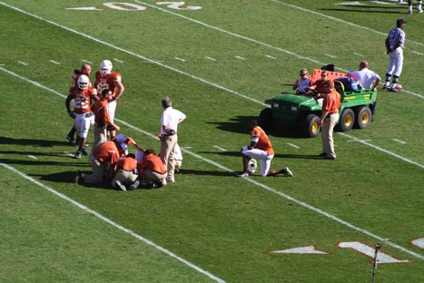
The retina holds a vast amount of information about the body and its overall health; it can help us detect signs of diabetes, high cholesterol, and brain tumors [3]. This is because the eye is the only region of the body that provides us with a clear view of arteries, veins, and cranial nerve. Now that we can provide retinal examinations on the go, can we definitively diagnose an athlete with a mild traumatic brain injury solely based on the microvasculature of the retina? According to Childs et al., “Mild traumatic brain injury (mTBI) accounts for 90% of all brain trauma. As only 15% of mTBI patients will have an identifiable intracranial lesion on brain computed tomography[4], injury is often considered as ‘insignificant’ when compared with the devastating and highly visible sequelae that follows severe TBI [5].” Furthermore, they also report, “arteriolar and venular tortuosity was significantly increased after mTBI. These findings remained consistent after additional adjustment for age in analysis of covariance model.” With more scientific evidence, I believe that ultrafast retinal examinations on the sidelines will one day revolutionize the way we diagnose traumatic brain injuries in high-profile sports leagues—a problem that has notoriously plagued professional sports.
In addition, are there other correlations we can begin to discover now that we can bring the eye clinic to the patient at the place of injury? Does the retina hold even more information than we previously thought? These are the types of questions we can begin to answer using Peek’s revolutionary product.
1. http://www.who.int/blindness/GLOBALDATAFINALforweb.pdf
2. http://www.designboom.com/design/eye-examination-kit-peek-retina-2015-index-award-08-28-2015/
3. http://www.webmd.com/eye-health/eye-tests-exams#
4. Jagoda AS. Mild traumatic brain injury: key decisions in acute management. Psychiatr Clin North Am 2010; 33:797–806
5. Childs C, Ong YT, Zu MM, Aung PW, Cheung CY, Kuan WS. Retinal imaging: a first report of the retinal microvasculature in
acute mild traumatic brain injury. European Journal of Emergency Medicine. 2014;21(5):388-389.
doi:10.1097/MEJ.0000000000000169.
Dor Shoshan is an MS1 at The University of Arizona College of Medicine – Phoenix. He graduated from Chapman University in 2015 with a Bachelor of Science in biological sciences. He is passionate about the intersection of science and technology with medicine—in particular, the way technology will shape the future of medicine and its perils. For recommendations about future articles, comments or questions, please do not hesitate to contact at shoshan[at]email.arizona.edu

